Isabella Coscarella
Flow of Life
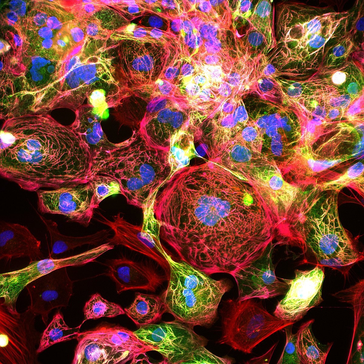
This is an image of human induced pluripotent stem cells (hiPSC) cardiomyocytes. These cells were reprogrammed into stem cells using CRISPR-Cas9 technology, and mutations associated with cardiomyopathy were introduced. Cardiomyopathies are diseases of the heart muscle in which the heart loses its ability to pump blood effectively. Since heart diseases are the leading cause of death in the United States of America, it is crucial to research the major causes of the pathology. Cardiomyopathies can be genetically inherited or acquired throughout life. Acquired cardiomyopathy can be a result of viral infections, autoimmune diseases, infiltrative disorders or inflammatory factors, and there is no cure as of today. Cardiomyopathies can trigger a variety of different alterations in morphology and function of proteins related to the contractility of the heart and therefore the heart’s performance. Then, it is important to investigate the functionality of proteins and its presence (or expression) in certain conditions. In the image, marked proteins that exert mechanical forces, such as myosin (green) and troponin (purple), show their expression, checking the presence of contractile apparatus of these cardiac cells.
Over the years, scientists have tried to find ways to target these proteins and develop specific medication that would act directly on them, helping the heart pump properly. If these specific targeted proteins are treated and start working correctly again, the muscle recovers its pumping mechanics. With the heart working properly, a patient originally diagnosed with cardiomyopathy can now live normally and have a high quality of life.
Artwork Description
This is an immunofluorescence microscopy of hiPSC (human-induced pluripotent stem cells) cardiomyocytes. The cardiomyocytes are treated with fluorescent markers to show specific proteins of interest. In the image, we have filamentous actin in red, ventricular myosin light chain in green, troponin T in purple, and the nucleus is dyed in blue using a common dye called DAPI. These cells must be cultured in favorable conditions for at least thirty days before an image can be acquired.